Filter
–
%
Erler-zimmer Right Ventricular Hypertrophy
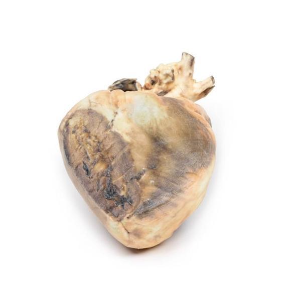
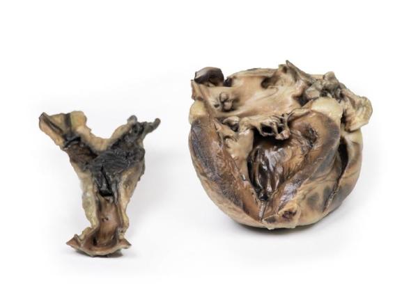
Produktinformationen "Right Ventricular Hypertrophy"Clinical HistoryThis 56-year old female suffered from emphysema and gave a 2-year history of increasing shortness of breath on exertion associated with recurrent attacks of bronchitis. On examination, she had a BP 160/90 mm Hg, pulse rate of 96 beats/min, and 6 cm of jugular venous congestion. The apex beat was impalpable, bilateral crepitations were heard and pitting oedema was present peripherally. Special investigations: ECG showed right heart strain pattern. Arterial blood examination showed respiratory acidosis. Despite treatment there was steady deterioration and death.Pathology The specimen is of the external surface of the heart viewed from the anterior aspect. The right ventricle is greatly enlarged and hypertrophied. All appears to be normal otherwise. This is an example of right ventricular hypertrophy (RVH) in a patient with emphysema.Further informationRVH usually occurs due to chronic lung disease or structural defects in the heart. One of the most common causes of RVH is pulmonary hypertension (PH), which leads to increased pulmonary artery pressure. As the right ventricle tries to compensate for this increased pressure it changes its shape and size causing hypertrophy and right ventricular wall thickness. The global incidence of PH is 4 per 1M people: RVH occurs in approximately 30 of these cases. Common causes of PH include chronic obstructive pulmonary disease (COPD), pulmonary embolism, and other restrictive lung diseases. RVH also occurs in response to structural defects in the heart, such as tricuspid insufficiency, which allows the backward flow of blood into the ventricle. Other structural defects that lead to RVH include tetralogy of Fallot, ventricular septal defects, pulmonary valve stenosis, and atrial septal defects. RVH is also associated with abdominal obesity and high systolic blood pressure.Weiterführende Links zu "Right Ventricular Hypertrophy" Fragen zum Artikel? Weitere Artikel von Nicht angegeben
Ab 847,28 €* 864,57 €* (2% gespart)
%
Erler-zimmer Tuberculosis
Produktinformationen "Tuberculosis"Clinical HistoryA 37-year old female presents with increasing thoracic back pain. She has a history of untreated human immunodeficiency virus (HIV) infection and pulmonary tuberculosis. History revealed ongoing low-grade fevers, chills and weight loss. Examination revealed a cachexic patient with tender thoracic vertebrae at multiple levels. Blood test showed an elevated serum calcium and erythrocyte sedimentation rate. X-ray of her spine showed lytic areas in the thoracic vertebrae. During her hospital admission, she developed urosepsis and died.Pathology The specimen is a portion of the patient’s thoracic vertebral column that has been sawn longitudinally and mounted to display the cut surface of 7 thoracic vertebrae. In all vertebrae, there are osteolytic areas, varying from 1 to 12 mm in diameter, which contain caseous degenerative material* (mostly now lost) and are surrounded by a thin zone of dense bone. The tuberculous inflammatory process has extended into one of the intervertebral discs, and has also spread outside the vertebral bodies to form collections of caseous material beneath the anterior longitudinal ligament. This is an example of tuberculous mycobacterial osteomyelitis of the vertebral column with paravertebral extension, also known as Pott’s Disease.Further InformationTuberculosis (TB) is a chronic pulmonary and systemic infectious disease caused by Mycobacteria tuberculosis. Transmission most commonly occurs via inhalation of aerosolized droplets of M. tuberculosis. Risk factors for contracting TB include being an inhabitant of a developing’ country where the disease may be endemic, immunosuppression (e.g. HIV, steroid use, anti-TNF use and diabetes), chronic lung disease (e.g. silicosis), alcoholism, and generalized malnutrition.After initial pulmonary infection of M. tuberculosis clinical manifestation varies. In 90 of individuals with an intact immune system, they enter an asymptomatic latent infection phase. This latent TB may reactivate at any time in the patients life. In the other 10 of patients, especially in the immunocompromised population, they develop primary disease, which is immediate active TB infection. Manifestations of primary TB include pulmonary infection symptoms (e.g. consolidation, effusion and hilar adenopathy) and extra pulmonary symptoms - lymphadenopathy, meningitis and disseminated miliary TB. Secondary tuberculosis occurs when there is reactivation of a previous latent TB infection. Around 10 of latent TB will reactivate usually during periods of weakened host immunity. Typical symptoms of reactivation are cough, haemoptysis, low grade fever, night sweats and weight loss.Osseous infection occurs 1-3 of patients with TB infection. There is a higher incidence of developing bone disease in patients from developing countries and immunocompromised patients. The TB usually spreads haematogenously from the site of active disease. Pott’s disease accounts for 40 of TB bone infections. The infection is destructive eroding vertebral discs and vertebrae leading to compression fractures, which may cause symptoms of cord or nerve root compression. Symptoms include pain at the site of disease, fevers, chills, weight loss, symptoms of compression and spinal deformities, such as kyphosis and scoliosis.TB diagnosis is usually made with a clinical history and chest x-ray and multiple sputum cultures. Mantoux skin tuberculin test and serum interferon gamma release assay may also be used to help screen for infection. Biopsies may be taken of suspected infection site for culture to assist diagnosis.Treatment involves prolonged courses of multiple antibiotics, which depend on the antibiotic resistance of the infecting mycobacterium species.* Caseous degeneration or necrosis is a unique form of cell death in which the tissue maintains a cheese-like appearance.Weiterführende Links zu "Tuberculosis" Fragen zum Artikel? Weitere Artikel von Nicht angegeben
Ab 202,49 €* 206,62 €* (2% gespart)
%
Erler-zimmer Circulus arteriosus Willisii
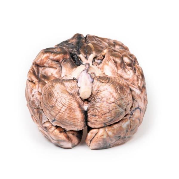
Produktinformationen "Circulus arteriosus Willisii"Dieses 3D gedruckte Präparat zeigt die intrakraniellen Arterien, die das Gehirn in Relation zu den unteren Anteilen des Gesichts- und Gehirnschädels versorgen. Dieser Druck entstand auf Grundlage sorgfältiger Segmentierung der angiographischen Daten. Das Modell zeigt die gepaarten Wirbelarterien, die durch das Foramen magnum in die Schädelhöhle eindringen und sich zur A. basilaris verbinden. Die Teilung der Arteria basilaris in ihre terminalen Zweige der A. cerebri posterior ist hier einsehbar. Die oberen Kleinhirnarterien entstehen knapp proximal zu dieser Verzweigung aus der A. basilaris. Detaillierte anatomische Beschreibung auf Anfrage.Weiterführende Links zu "Circulus arteriosus Willisii" Fragen zum Artikel? Weitere Artikel von Erler-Zimmer
Ab 1.513,57 €* 1.544,45 €* (2% gespart)
%
Erler-zimmer Unterarm und Hand oberflächliche und tiefe Präparation
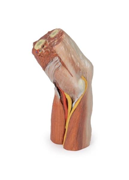
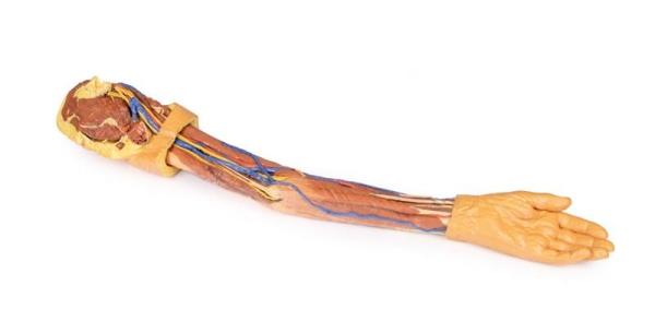
Produktinformationen "Unterarm und Hand oberflächliche und tiefe Präparation"Dieses 3D-gedruckte Präparat zeigt eine sowohl oberflächliche als auch tiefe Präparation der anterioren Seite eines rechten distalen Arms, Unterarms und Hand.Detaillierte anatomische Beschreibung auf Anfrage.Weiterführende Links zu "Unterarm und Hand oberflächliche und tiefe Präparation" Fragen zum Artikel? Weitere Artikel von Erler-Zimmer
Ab 1.298,64 €* 1.325,14 €* (2% gespart)
%
Erler-zimmer Tiefe Gesichtsknochen/Fossa infratemporalis
Produktinformationen "Tiefe Gesichtsknochen/Fossa infratemporalis"In diesem 3D gedruckten Präparat einer mittelsagittal geschnittenen rechten Gesichts- und Halshälfte wurden Ramus, Processus coronoideus mandibulae und Mandibulakopf entfernt, um den tiefen Teil der Fossa infratemporalis freizulegen. Die seitlichen Flügelmuskeln (M. pterygoideus lateralis) wurden ebenfalls entfernt, um das laterale Pterygotid und die hintere Oberfläche des Oberkiefers freizulegen. Der Musculus buccinator wurde belassen, und seine Ansätze können von der äußeren Seite des Oberkiefers, der Raphe pterygomandibularis und der Außenseite des (zahnlosen) Unterkiefers betrachtet werden. Detaillierte anatomische Beschreibung auf Anfrage.Weiterführende Links zu "Tiefe Gesichtsknochen/Fossa infratemporalis" Fragen zum Artikel? Weitere Artikel von Erler-Zimmer
Ab 519,23 €* 529,82 €* (2% gespart)
%
Erler-zimmer Sinus Pathways
Produktinformationen "Sinus Pathways"This 3D model provides a midsagittal to parasagittal segment of a right head to demonstrate the relationships and passageways of the paranasal sinuses. These passageways have been highlighted with thin coloured markers to indicate the relationship of these communicating routes between the paranasal sinuses and the nasal cavity.Starting anteriorly in the nasal cavity, the opening of the nasolacrimal duct (white) is present just deep to the inferior nasal conchae. The middle nasal concha has been sectioned to allow for a clear view of the opening of the maxillary sinus (visible in the parasagittal plane) across the semilunar hiatus (green), as well as the drainage of the frontal sinus (blue with the sinus visible superiorly in the section and in the transverse cut through the specimen) and the anterior (orange) and middle (yellow) ethmoidal cells. The opening of the posterior ethmoidal cells into the superior meatus is shown through the purple marker, which is visible within a small opened window into the ethmoid just superior to the nasal cavity. Finally, the opening of the sphenoid sinus is marked in red and visible through the opened sphenoid sinus itself just superior to the nasopharyngeal region.In addition to these pathways, this 3D model also captures some of the surrounding anatomy within the section. Visible in the midsagittal view are the other primary structures of the nasal cavity from the nostril to the opening of the auditory tube posteriorly. The soft palate and uvula are preserved, as is the rest of the pharynx just to the level of the epiglottis and collapsed laryngeal region at the inferior part of the preserved specimen. The oral cavity is displayed in cross section, with distinct genioglossus and geniohyoid muscles. In the cranial cavity, parts of the brain are preserved including the inferior parts of the frontal lobe and the right optic nerve/chiasm/tract. The pituitary gland is visible in cross-section just superior to the sphenoid sinus. The pons, medulla oblongata, and most of the cerebellum are present, with a small part of the tentorium cerebelli separating the cerebellum from the right occipital lobe of the cerebrum. On the parasagittal side of the specimen there is a continuation of the tentorium cerebelli separating these parts of the brain, with clear cross-sections of the transverse sinus and part of the sigmoid sinus on either side of the cerebellum. Overlying this is a small part of the medial temporal lobe of the cerebellum with part of the anterior horn of the lateral ventricle deep within the lobe.Weiterführende Links zu "Sinus Pathways" Fragen zum Artikel? Weitere Artikel von Nicht angegeben
Ab 813,35 €* 829,94 €* (2% gespart)
%
Erler-zimmer Atrial septal defect
Produktinformationen "Atrial septal defect"Clinical HistoryA 10-year-old girl with a known congenital heart was admitted for surgical repair because of the recent onset of cyanosis and cardiac failure. On examination, she was breathless with a blood pressure of 105/60mm/Hg and a pulse rate of 140/min. There was a loud heart murmur in the fourth left intercostal space adjacent to the sternum. The jugular venous pressure was elevated, and there were bilateral pulmonary basal crepitations but no peripheral oedema. At operation, the defect was repaired however, death followed a sudden post-operative deterioration of unknown cause.Pathology The heart is viewed from the left side. The left atrium has been opened to display a large ovoid defect 3.5 cm in greatest diameter in the inter-atrial septum. Only a small postero-inferior crescentic rim of septum remains. The left ventricle is small, and the right ventricle is hypertrophied (see posterior aspect of specimen where part of the right postero-lateral wall of the right ventricle has been cut away to demonstrate the thickened wall). The pulmonary artery, seen to the left of the atrial cavities, is greatly enlarged. The smaller vessel seen lying above the cut end of the pulmonary artery is the aortic arch. The cut edge of a lumen 8 mm in diameter immediately below the cut end of the pulmonary artery is the left auricular appendage.Further InformationAtrial septal defect is usually asymptomatic early in life, even when large. Symptoms may not develop until adult life. The onset of symptoms is due to reversal of the initial left-to-right shunt as a result of increasing right ventricular hypertrophy and pulmonary hypertension. The ensuing right-to-left shunt is associated with cyanosis and dyspnoea, and ultimately leads to congestive cardiac failure.There are several types of atrial septal defects, including:Secundum - This is the most common type of ASD and occurs in the middle of the wall between the atria (atrial septum). Primum - This defect occurs in the lower part of the atrial septum and might occur with other congenital heart problems.Sinus venosus - This rare defect usually occurs in the upper part of the atrial septum and is often associated with other congenital heart problems.Coronary sinus - In this rare defect, part of the wall between the coronary sinus which is part of the vein system of the heart and the left atrium is missing.It is not known why all atrial septal defects occur, but some congenital heart defects appear to be familial and sometimes occur with other genetic problems, such as Trisomy 21 (Down’s syndrome). Some conditions during pregnancy can increase the risk of having a baby with a heart defect, including acute infections such as Rubella infection drug, tobacco or alcohol use, or exposure to certain substances (such as cocaine) during the first trimester of pregnancy and underlying systemic conditions, such as diabetes or systemic lupus erythematosus.Weiterführende Links zu "Atrial septal defect" Fragen zum Artikel? Weitere Artikel von Nicht angegeben
Ab 1.039,59 €* 1.060,80 €* (2% gespart)
%
Erler-zimmer Abdomen vasculature
Produktinformationen "Abdomen vasculature"Coeliac TrunkSupplying the embryological foregut, the celiac trunk arises from T12 spinal level. Branches that can be observed in this specimen include the Left gastric artery arising from the left portion of the celiac trunk remains of the splenic artery arising from the celiac trunk and visible passing to the left hypochondrium the Common hepatic artery, located to the right of the celiac trunk and giving off key branches the Gastroduodenal artery, branching inferior to into the right gastric artery, and provide an anastomosis to the superior mesentery artery via the superior pancreaticoduodenal and the Proper hepatic artery, beginning after the gastroduodenal artery, branching to form the Left hepatic artery, the first branch of the proper hepatic artery, Right hepatic artery, located inferiorly, eventually giving rise to the Cystic artery, connecting to the gallbladder.Superior Mesenteric Artery and Inferior Mesenteric ArterySupplying the midgut and hindgut respectively, the superior mesenteric and inferior mesenteric artery arise at the L1 and L3 vertebral levels, respectively.While both have key branches, this specimen does not preserve them in their entirety. The Superior mesenteric artery can be seen in the model exiting below the pancreas, dividing out into many branches and the Inferior mesenteric artery can be observed descending on the left of the abdominal aorta. The left colic artery, moving laterally, can be seen leaving the IMA to give rise to the marginal arteries that supply to the colon.Venous System of the AbdomenThe superior mesenteric vein can be seen posterior to the superior mesenteric artery, notably less tubular than its arterial counterpart.In the specimen, the left anatomical lobe of the liver has been removed, exposing portal vein branches. These will supply nutrients from the gastrointestinal system to the hepatocytes which will then connect back to the venous system through the hepatic veins. This will then meet the Inferior Vena Cava.Hilum of the KidneyThe right kidney shows typical anatomy, as opposed to the left kidney which shows anatomical variation. Seen at the right kidney are the Right renal vein, most superior, merging directly into the IVC, the Right renal artery, most inferior, passing deep to the IVC from its origin from the abdominal aorta and the Right ureter, coursing superficial to the right renal artery to eventually travel inferiorly.The left kidney presents unique variation at the hilum with key structures as follows. The Left renal vein, most inferior (as opposed to the usual superior) and is highly subdivided. The Left renal artery, most superior (as opposed to the usual inferior) and the Left ureter, can be seen descending from the hilum and medial to the kidney.Muscles, Nerves and Other VasculatureThe psoas major and iliacus muscle can be seen on both sides of the specimen and surrounding them, key branches of the lumbar plexus can be seen, particularly on the left side. The Iliohypogastric nerve, continuing laterally as the most superior of the nerves present and the Ilioinguinal nerve, inferior to the iliohypogastric, directed towards the inguinal canal. The Femoral nerve, originating deep to and entering view lateral to psoas major and the Genitofemoral nerve, coursing superficial to psoas major, dividing into the genital and femoral branches of innervation.Medial to the psoas major, the left testicular artery and left testicular vein can be seen (as this is a male specimen). While the artery will receive blood directly from the aorta, the left testicular vein will drain to the left renal vein.The right sided testicular vasculature can also be observed however the right testicular vein drains directly into the IVC.The branch of the iliolumbar artery that anastomoses with the iliac circumflex artery can be observed passing under the testicular artery and vein and under the ureter.GallbladderJust inferior to the liver, the gallbladder can be observed with the cystic artery moving inferiorly to meet it. The cystic duct can also be seen moving from the gallbladder, meeting the common hepatic duct moving from the liver to form the common bile duct.Weiterführende Links zu "Abdomen vasculature" Fragen zum Artikel? Weitere Artikel von Nicht angegeben
Ab 5.802,00 €* 5.920,40 €* (2% gespart)
%
Erler-zimmer Fuß Strukturen der Plantarfläche
Produktinformationen "Fuß Strukturen der Plantarfläche"Dieses 3D gedruckte Präparat stellt die Anatomie des linken distalen Unterschenkels und die tiefen Strukturen der Plantarfläche des Fußes dar. An der kranialen Schnittfläche lassen sich Tibia, Fibula, interossäre Membran und Beinmuskulatur erkennen. Medial, auf Höhe des Sprunggelenks, sind die langen Sehnen der Dorsalextensoren und der Plantarflexoren oberhalb des Kapsel-Bandapparates und der extrakapsulären Bänder sichtbar. Detaillierte anatomische Beschreibung auf Anfrage.Weiterführende Links zu "Fuß Strukturen der Plantarfläche" Fragen zum Artikel? Weitere Artikel von Erler-Zimmer
Ab 1.057,69 €* 1.079,27 €* (2% gespart)
%
Erler-zimmer Carcinoma of Larynx
Produktinformationen "Carcinoma of Larynx"Clinical HistoryA 47-year-old male presents with a 13-month history of dysphonia and odynophagia at the level of his thyroid cartilage. He has a significant smoking history. Investigations revealed a laryngeal tumour. He received radiotherapy to the tumour followed by a laryngectomy. Six months later pulmonary metastases were discovered, and he subsequently died.Pathology This is the patient’s laryngectomy specimen. The larynx has been sliced open and is viewed from the posterior aspect. There is significant right vocal cord distortion by an irregular ulcerating tumour. Mucosal congestion is also noted. Histologically this was a well differentiated squamous cell carcinoma (SCC).Further InformationOver 95 of laryngeal cancers are SCC. The tumour usually develops on the vocal cords but may occur above or below the cords, on the epiglottis, aryepiglottic folds or in the pyriform sinuses. The cancer usually begins as carcinoma in situ, progressing to ulcerated and fungating carcinoma with continued exposure to carcinogens.The greatest risk factors for developing cancer of the larynx are tobacco smoke and alcohol consumption. Human Papilloma Virus (HPV) infection, asbestos exposure and irradiation have also shown increased incidence. Males are affected more than females. It most frequently presents in the 6th decade of life.Laryngeal cancer may spread by invading into surrounding structures, via lymphatics usually to local cervical nodes or haematogenous metastasis most commonly to the lungs. Common symptoms on presentation include dysphonia, dysphagia, odynophagia, globus and cough. Less commonly haemoptysis, stridor, dyspnoea and halitosis may be described. Treatment varies on the stage of the disease. Smoking and alcohol cessation are important for all disease stages. In early disease laryngeal preservation treatments may include laser therapy, microsurgery and radiotherapy. Later stage disease treatments may involve a combination of laryngectomy, radiotherapy and chemotherapy.Weiterführende Links zu "Carcinoma of Larynx" Fragen zum Artikel? Weitere Artikel von Nicht angegeben
Ab 303,16 €* 309,35 €* (2% gespart)
%
Erler-zimmer Transverse Section of the head
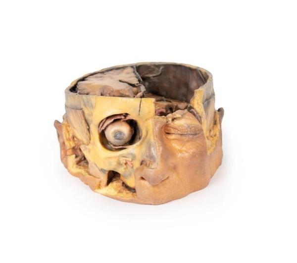
Produktinformationen "Transverse Section of the head"This 3D model preserves a transverse section through the cranial cavity with partial dissection of the brain and exposure of the left orbital roof, alongside a deep dissection of the face and temporomandibular joint region.Within the cranial cavity, the dura mater has been largely removed from the anterior cranial fossa, with retention of the layer in part across the middle and posterior cranial fossae. On the right side, the cerebrum has been dissected to expose the lateral ventricle and to open the lateral fissure to demonstrate the course of the middle cerebral artery between the frontal, parietal and temporal lobes. A more significant dissection of the brain on the left side allows for an appreciation of the midline third ventricle and retained septum pellucidum on the right side, the falx cerebri (with the superior sagittal sinus visible in cross-section), and parts of the anterior and posterior horns of the lateral ventricle with choroid plexus. This differential dissection of the brain also provides an excellent view of the optic nerves, chiasm and tracts, and the relation of these nervous structures to the left internal carotid artery, and bases of the anterior and middle cerebral arteries.Anteriorly in the cranial cavity, the left optic nerve can be followed into the left orbit, which has been opened to expose several key orbital structures. Centrally, the frontal nerve is well-preserved on the levator palpebrae superioris muscle. Laterally, the lacrimal gland rests in the superior quadrant, while medially the partial dissection into the frontal bone and sinuses affords a clear view of the superior oblique muscle passing through the trochlea. Deep to these superficial structures extraocular fat has been removed to show the medial rectus muscle laterally, and the nasocilliary nerve and medial rectus muscles medially.In the face, the skin, superficial tissue, orbicularis oculi and extraocular fat have been removed from the right orbit to expose the extraocular muscles and lacrimal gland. The levator palpebrae superioris is well-defined despite being detached from the superior tarsal plate. The reflection of the superior oblique muscle from the trochlea onto the eye can be seen, as well as the insertions of the medial and lateral rectus muscle, and the full course of the inferior oblique.Across the rest of the right side of the face and temporal region a deep dissection has exposed a number of structures. Inferior to the orbital margin, the infraorbital artery and nerve have been exposed exiting via the infraorbital foramen. The superficial and deep heads of the masseter are well defined, and the partial dissection of the temporalis muscle provides a perspective on its broad origin and depth of fibres near pterion (and in contrast to the exposed and undissected right side). Inferior to the zygomatic, the parotid gland has been dissected to expose the mandibular condyle resting in the glenoid fossa and to demonstrate the relationship of the external ear relative to the external auditory meatus.Weiterführende Links zu "Transverse Section of the head" Fragen zum Artikel? Weitere Artikel von Nicht angegeben
Ab 2.177,59 €* 2.222,03 €* (2% gespart)
%
Erler-zimmer LungCystic Fibrosis
Produktinformationen "LungCystic Fibrosis"Clinical HistoryA 6-year old girl was admitted with a productive cough, dyspnoea and fevers. She became increasingly hypotensive and dies soon after admission. She had a previous history of recurrent pneumonia and meconium ileus. The clinical diagnosis was cystic fibrosis (mucoviscoidosis). Her sister died aged 3 from the same disease.Pathology The lung parenchyma shows extensive changes mainly with a bronchial distribution. Many bronchi are dilated (bronchiectasis) and contain thick, yellowish, purulent material. These changes are most marked in the upper lobe, at the apex of which a small focus of honeycomb’ change is also seen. Multiple abscesses are present, especially in the basal and central parts of the lower lobe. The base of the lower lobe is severely affected with fibrosis and consolidation being evident. There is very little normal lung tissue remaining. These pathological changes are characteristic but not pathognomonic of cystic fibrosis.Further InformationCystic fibrosis (CF) is an inherited disorder of chloride ion transport. Mutations in the cystic fibrosis conductance regulator (CFTR) gene on chromosome 7 cause defects in the chloride channel protein leading to dysfunction of the chloride channels. This causes increased water absorption in exocrine glands and epithelium of the respiratory, gastrointestinal and reproductive tracts. These dehydrated viscous secretions then obstruct these organ passage causing clinical features including: persistent pulmonary infection, pancreatic insufficiency, liver cirrhosis, intestinal obstruction, male infertility, and elevated sweat chloride levels. In the airway, CF patients have decreased chloride secretion and increased water reabsorption. This causes dehydrated mucous lining the airways leading to defective mucociliary action, mucous obstructing the airway, bronchiole dilatation (bronchiectasis) and secondary infection. Staphylococcus aureus, Haemophilus influenzae and Pseudomonas are the most common bacteria causing CF patients’ lower respiratory tract infections. Chronic bronchitis and bronchiectasis develops as a result. Pulmonary issues are the highest cause of mortality in CF patients. The average life expectancy is between 40-50 years of age in developed countries.CF occurs in around 1 in 3000 live births. It is inherited in an autosomal recessive pattern. It is most common in fair-skinned populations: with 1 in 20 being a carrier of the gene. Symptoms can present in-utero or even up to adolescence, depending on the severity of the disease. It is now most commonly diagnosed with the neonatal screening test for immunoreactive trypsinogen (a pancreas enzyme precursor). If this screening test is positive, a formal diagnosis is made with a sweat test showing >60mmol/L of chloride.Weiterführende Links zu "LungCystic Fibrosis" Fragen zum Artikel? Weitere Artikel von Nicht angegeben
Ab 259,05 €* 264,33 €* (2% gespart)
%
Erler-zimmer Astrocytoma
Produktinformationen "Astrocytoma"Clinical HistoryA 73-year-old female was admitted with new left-sided hemiplegia. On further questioning she revealed a 3-month history of headaches, nausea and deteriorating balance. CT brain revealed an inoperable brain tumour. She died 1 week after being admitted.PathologyThis brain specimen is a coronal section. In the right temporal lobe, a poorly demarcated tumour is present. There is enlargement of the hemispheres and flattening of the gyral pattern. From the posterior aspect of the specimen subfalcine herniation* is appreciated and the tumour appear less well differentiated with haemorrhagic and necrotic foci. Histology of this tumour showed an astrocytoma, Grade III/IV.*In subfalcine (or cingulate) herniation, the most common type of brain herniation, the innermost part of the frontal lobe is pushed under part of the falx cerebri, between the two hemispheres of the brain.Further InformationGliomas are the second most common cancer of the central nervous system after meningiomas. The term glioma refers to tumours that are histologically similar to normal glial cells i.e. astrocytes, oligodendrocytes and ependymal cells. They arise from a progenitor cell that differentiates down one of the cell lines. Astrocytomas develop from the astrocyte lineage of glial cells. Tumours are staged according to histological differentiation and range from diffuse astrocytoma (Grade II/IV) to anaplastic astrocytoma (Grade III/IV) to glioblastoma (Grade IV). Histological features include the prominent eosinophilic cytoplasm in some astrocytic tumour cells (gemistocytes) as well as a fibrillary background.Astrocytomas occur most commonly between the fourth and sixth decades of life. Tumours usually occur in the cerebral hemispheres but may also occur in the cerebellum, brainstem or spinal cord. They most commonly present with seizures, headaches, nausea and focal neurological deficits depending on area involved. Without treatment Grade III median survival is 18 months. Treatment includes surgical resection, radiotherapy, chemotherapy or a combination thereof, depending on the clinical context.Weiterführende Links zu "Astrocytoma" Fragen zum Artikel? Weitere Artikel von Nicht angegeben
Ab 259,05 €* 264,33 €* (2% gespart)
%
Erler-zimmer Oberflächliche Venen der unteren Extremität
Produktinformationen "Oberflächliche Venen der unteren Extremität"Dieses 3D gedruckte Präparat zeigt eine oberflächliche Präparation der linken unteren Extremität, von knapp kranial des Kniegelenks bis zu dem kompletten Fuß. Die Haut und die oberflächliche Faszie wurden entfernt, um die oberflächlichen Venenformationen des Unterschenkels einschließlich des Venengeflechts des Fußrückens, der Vena saphena magna (einschließlich zahlreicher Zuflüsse) und der Vena saphena parva (einschließlich zahlreicher Zuflüsse) auf der Fascia cruris darzustellen.Detaillierte anatomische Beschreibung auf Anfrage.Weiterführende Links zu "Oberflächliche Venen der unteren Extremität" Fragen zum Artikel? Weitere Artikel von Erler-Zimmer
Ab 2.542,97 €* 2.594,87 €* (2% gespart)
%
Erler-zimmer Left cerebral infarct
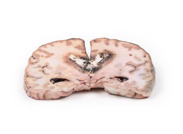
Produktinformationen "Left cerebral infarct"Clinical HistoryThe patient was a 51-year old woman who had a cerebro-vascular accident resulting in a left hemiplegia 2 years prior to death. At necropsy, she had severe generalized atherosclerosis and an old left ventricular myocardial infarct with an overlying mural thrombus.PathologyA coronal section of the cerebral hemispheres shows irregular cystic cavities in the territory of distribution of the right middle cerebral artery. The cavities of the infarct have irregular, yellow walls and show partial collapse. There is compensatory dilatation of the left lateral ventricle. On the posterior aspect, the arteries below the mammillary bodies were moderately atheromatous, although this is difficult to visualise macroscopically.Further InformationBecause of the underlying history of myocardial disease with the presence of the mural thrombus, it is assumed that her cerebral infarct was probably caused by a thromboembolus.Weiterführende Links zu "Left cerebral infarct" Fragen zum Artikel? Weitere Artikel von Nicht angegeben
Ab 247,74 €* 252,79 €* (2% gespart)
%
Erler-zimmer Liver with vessels and gallbladder
Produktinformationen "Liver with vessels and gallbladder"The size and shape of this specimen varies somewhat from a typical liver. It is less wedge-shaped and longer in the superoinferior dimension (on the posterior view this would translate to a greater vertical height). Normally, a liver is less than 16cm in the midclavicular line.1 This specimen measures approximately 18cm in the midclavicular line, suggesting some degree of hepatomegaly. However, it is worth mentioning that some measurement distortion may have occurred based on the fixing and curation of the specimen and it must be noted that the accuracy of estimating liver size using a single parameter is limited. Liver measurements diagnostic of hepatomegaly vary depending on normal anatomical variation in liver size and morphology, the method of measurement, and patient features such as sex and BMI.An alternative explanation could be normal anatomical variation. However, this specimen does not fit the description of the most common anatomical variations confused with hepatomegaly Riedel’s lobe (a downward projection of the right lobe), beaver tail’ liver (an elongated left lobe), or a papillary process projecting from the caudate lobe.Weiterführende Links zu "Liver with vessels and gallbladder" Fragen zum Artikel? Weitere Artikel von Nicht angegeben
Ab 1.864,24 €* 1.902,29 €* (2% gespart)
%
Erler-zimmer Carcinoma of Larynx
Produktinformationen "Carcinoma of Larynx"Clinical HistoryA 74-year old male presented with a 2-months history of dysphagia, dysphonia and weight loss. He had a history of heavy alcohol consumption and smoked 40 cigarettes per day for 40 years. Investigation discovered a laryngeal tumour. He received radiotherapy but his tumour reoccurred. He died 9 months after his initial presentation.PathologyThe specimen consists of tongue, pharynx, larynx, oesophagus and trachea and has been mounted in the coronal plane. The oesophagus and trachea have been opened from the posterior aspect. There is a 5 x 4 x 2 cm fungating carcinoma evident extending into both pyriform fossae. The surface of the tumour is irregular with shaggy areas of necrosis. The tumour has arisen from the larynx and involves both vocal cords, the left aryepiglottic fold and both pyriform fossae.Further InformationOver 95 of laryngeal cancers are squamous cell carcinomas. The tumour usually develops on the vocal cords but may occur above or below the cords, on the epiglottis, aryepiglottic folds or in the pyriform fossae. The cancer usually begins as squamous cell carcinoma in situ, progressing to ulcerated and fungating invasive carcinoma with continued exposure to carcinogens. The greatest risk factors for developing cancer of the larynx are tobacco smoke and alcohol consumption. Human Papilloma Virus (HPV) infection, asbestos exposure and irradiation have also been shown to be associated with increased incidence of head-and-neck squamous cell carcinoma (HNSCC). Males are affected more than females. It most frequently presents in the 6th decade of life. Laryngeal cancer may spread by invading into surrounding structures, via lymphatics usually to local cervical nodes, or haematogenous metastasis most commonly to the lungs. Common symptoms of HNSCC on presentation include dysphonia, dysphagia, odynophagia, globus and cough. Less commonly haemoptysis, stridor, dyspnoea and halitosis may be described.Treatment varies on the stage of the disease. Smoking and alcohol cessation are important for all disease stages. In early disease laryngeal preservation treatments may include laser therapy, microsurgery and radiotherapy. Later stage disease treatments may involve a combination of laryngectomy, radiotherapy and chemotherapy.HPV-related HNSCC have better outcomes than those non-HPV positive tumours. HPV-vaccination programmes have been introduced in several countries, including Australia and the UK, for both boys and girls, in order to reduce their risk for HNSCC.Weiterführende Links zu "Carcinoma of Larynx" Fragen zum Artikel? Weitere Artikel von Nicht angegeben
Ab 661,76 €* 675,27 €* (2% gespart)
%
Erler-zimmer Lung Slab, Hilum removed
Produktinformationen "Lung Slab, Hilum removed"The lung has been dissected following a parasagittal plane, removing the mediastinal surface. Ordinarily, the pulmonary arteries, veins and bronchi can be observed entering the lung in the hilum but the primary bronchi cannot be seen in this specimen as they have already divided substantially. It is unclear how far laterally the specimen has been dissected hence the bronchi subdivision level (secondary or tertiary) cannot be determined.The cardiac impression is formed by the left ventricle of the heart resting on the mediastinal surface of the lung. Although the lung has been dissected following a parasagittal plane, the cardiac impression can still be observed as it is the most concave area of the medial surface of the lung.The lung sits above the diaphragm, forming the concave diaphragmatic surface. The pleura has not been preserved in this specimen, but ordinarily, there exists a diaphragmatic recess bounded by the costal and diaphragmatic pleura. This would lie between the lung’s diaphragmatic impression and the diaphragm.Weiterführende Links zu "Lung Slab, Hilum removed" Fragen zum Artikel? Weitere Artikel von Nicht angegeben
Ab 882,35 €* 900,35 €* (2% gespart)
%
Erler-zimmer Weibliches rechtes Becken
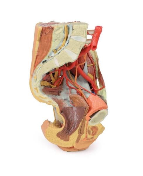
Produktinformationen "Weibliches rechtes Becken"Dieses 3D gedruckte Präparat stellt ein weibliches rechtes Becken dar, das entlang der mittelsagittalen Ebene und transversal auf Höhe des Wirbelkörpers L4 und des proximalen Oberschenkels geschnitten ist. Das Exemplar wurde so seziert, dass die tiefen Strukturen des großen und kleinen Beckens, der unteren vorderen Bauchdecke und des Leistenbereichs, des Femurdreiecks und des Gesäßbereichs veranschaulicht werden. Detaillierte anatomische Beschreibung auf Anfrage.Weiterführende Links zu "Weibliches rechtes Becken" Fragen zum Artikel? Weitere Artikel von Erler-Zimmer
Ab 2.713,78 €* 2.769,17 €* (2% gespart)
%
Erler-zimmer Weibliches linkes Becken und proximaler Oberschenkel
Produktinformationen "Weibliches linkes Becken und proximaler Oberschenkel"Dieses 3D gedruckte Präparat eines weiblichen linken Beckens und proximalen Oberschenkels in 3D-Druck zeigt sowohl oberflächliche als auch tiefe Strukturen des großen und kleinen Beckens, der Leistenregion, des Femurdreiecks und der Gesäßregion. Das Exemplar wurde quer durch den vierten Lendenwirbel geschnitten und zeigt den Querschnitt durch die Muskulatur (epaxiale Muskulatur, M. psoas und M. quadratus lumborum) und durch die Cauda equina im Wirbelkanal. Die ventralen und dorsalen Wurzeln der Cauda equina sind auch im Sagittalschnitt bei ihrem Austritt aus den intervertebralen und sakralen Foramina sichtbar. Detaillierte anatomische Beschreibung auf Anfrage.Weiterführende Links zu "Weibliches linkes Becken und proximaler Oberschenkel" Fragen zum Artikel? Weitere Artikel von Erler-Zimmer
Ab 2.702,47 €* 2.757,62 €* (2% gespart)
%
Erler-zimmer Intussusception of small bowel due to metastatic tumour
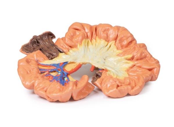
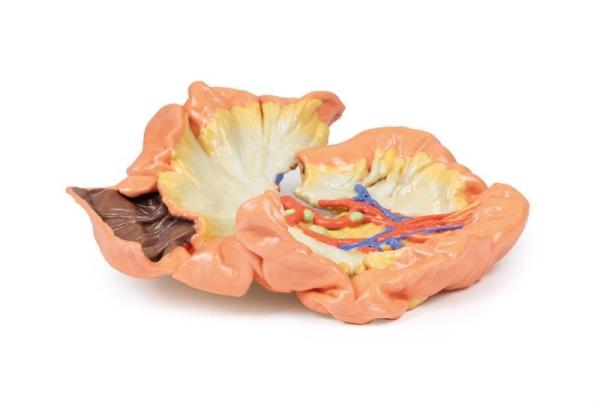
Produktinformationen "Intussusception of small bowel due to metastatic tumour"Clinical HistoryA 66-year-old woman suffered sudden onset of severe colicky central abdominal pain, somewhat relieved by drawing up her knees. She passed a stool containing mucus and blood ("like redcurrant jelly"). On examination, there was a mass in the left hypochondrium, which hardened with each spasm of pain. The specimen was resected at laparotomy.Pathology The specimen is a segment of small bowel, approximately 20 cm in length, with attached mesentery up to 2 cm in width (more evident on the uncut aspect of the specimen). About 5 cm from the proximal surgical resection margin (which is at the left hand of the specimen), a polypoid tumour 3 cm in diameter has become invaginated into the lumen of the bowel, and has been propelled distally, forming an intussusception 13 cm in length. The tumour is seen at the apex of the intussusception (near the right hand side of the specimen). The congestion and exudate seen on the mucosal surface of the intussusception (invaginated portion) are features considered with early ischaemic necrosis. The histological diagnosis is not recorded in this case however, the macroscopic appearance is consistent with a metastatic malignant tumour, although the possibility of a primary tumour cannot definitely be excluded.Further InformationIntussusception of the small bowel is most common in children, in whom it is usually due to invagination of swollen lymphoid tissue (Peyers patches) in the wall of the distal ileum. In adults, it is rare, causing only between 1 - 5 percent of cases of bowel obstruction. The usual cause a polypoid tumour, as seen in this specimen, acting as a pathological lead point being pulled forward by peristalsis, and thereby causing telescoping of the affected portion of bowel distally. Presentation may be of intermittent symptoms of bowel obstruction and in some cases excruciating pain. Classification of intussusception can be by causal pathology or by location. Abdominal CT scan will typically demonstrate a typical target sign with alternating hyper/hypodense layers.Weiterführende Links zu "Intussusception of small bowel due to metastatic tumour" Fragen zum Artikel? Weitere Artikel von Nicht angegeben
Ab 314,47 €* 320,90 €* (2% gespart)
%
Erler-zimmer TracheaHodgkin Lymphoma
Produktinformationen "TracheaHodgkin Lymphoma"Clinical HistoryA 45-year old male presented with a lump in his left supraclavicular area. The swelling had been increasing in size over 6 months. Excision biopsy of the lump showed Hodgkin lymphoma (HL). Ten months later he was readmitted with left shoulder pain and swelling of his left arm. Examination revealed generalised lymphadenopathy with significant swelling in his left supraclavicular and axillary regions. He was treated with radiotherapy and Thiotepa chemotherapy. He developed vomiting. A subsequent barium meal showed duodenal obstruction from extrinsic lymph node compression. He continued to deteriorate and died 2 weeks after readmission.Pathology The 3D print shows the tracheal bifurcation with adjacent para-tracheal and peri-bronchial lymph nodes. The trachea has been opened longitudinally and is viewed from behind. The para-tracheal lymph nodes are pale and matted (fused) together. Similar abnormal tissue is seen as a confluent pale mass on the left side of the trachea, above the aortic arch, which is seen cut in cross-section as a void space with branches arising. The peri-bronchial lymph nodes are also enlarged, and contain carbon pigment. The circumscribed small paler areas in the lymph nodes and extra-nodal tumour are foci of necrosis. There is an atheroma in the wall of the aorta but it is difficult to see in the 3D print.Further InformationHodgkin Lymphoma (HL) is a malignancy of lymphocytes. It is characterised by the presence of neoplastic giant cells called Reed Sternberg cells. There are 5 main subtypes according to the WHO Lymphoma Classification, based on the morphology, immunophenotyping and genetics. Activation of the transcription factor NF-kB is a common pathway of tumorigenesis among the subtypes. This promotes proliferation, reduces apoptosis, and induces expression of cytokines that recruit the immune cells that surround Reed Sternberg cells in HL.There is a bimodal age distribution with a peak in late adolescence/early adulthood and a second peak in older adults. HL accounts for just under 1 of all cancers worldwide. Infection with Epstein Barr Virus (EBV) contributes to the pathogenesis of the main subtypes of HL. The viral genome causes genetic alterations that lead to aberrant signal pathways, although the precise mechanism is not known. Immunosuppression (e.g. HIV infection or post- organ transplant) and positive HL family history are also risk factors. HL commonly presents as painless lymphadenopathy, pruritus, weight loss, fevers and night sweats. Later disease sees organ spread to the spleen, liver and bone marrow. Compressive symptoms can arise from enlargement of lymph nodes and infiltrated organs. HL is diagnosed with staging CT scan, excision biopsy of involved nodes and bone marrow biopsy. Treatment involves radiotherapy and chemotherapy. Although previously incurable, the overall survival of HL has improved significantly over the last 5 decades as a result of modern therapies: diagnosed at an early stage, it is almost 90, and even later stage disease has a favourable prognosis.Weiterführende Links zu "TracheaHodgkin Lymphoma" Fragen zum Artikel? Weitere Artikel von Nicht angegeben
Ab 259,05 €* 264,33 €* (2% gespart)
%
Erler-zimmer Osteosarcoma of femur
Produktinformationen "Osteosarcoma of femur"Clinical HistoryA 16-year old male presented with a 3-month history of increasingly swollen and painful right knee. On examination, there was a palpable tender swelling above the right knee. Blood test showed a raised Alkaline Phosphatase level. A knee x-ray showed periosteal reactive changes in the distal femur suspicious for a bone malignancy. The patient then underwent staging CT and MRI evaluation of the right leg. He underwent adjuvant chemotherapy prior to resection of his right femur. He made a full recovery.Pathology The specimen is the patient’s excised distal femur. On the cut surface, there is a large pale infiltrating tumour, 10 cm in greatest diameter, extending through the periosteum near the articular surface. This is an osteosarcoma of the femur.Further InformationOsteosarcomas are a malignant tumour of bones that are characterised by the production of osteoid matrix or immature bone. It is the most common primary malignancy of bone. Most occur in the distal femur with the tibia and humerus being the most frequent sites affected. Men are more commonly affected than women. They occur in a bimodal age distribution, with most occurring in children and adolescents under 20 years of age and the second peak occurring in older adults over 60.Secondary osteosarcomas are more common in older patients. Secondary osteosarcomas occur in patient’s bones with predisposing conditions such as Pagets disease, bone infarcts and previous irradiation. Mutations in tumour suppressors and oncogenes, such as RB, TP53 and INK4a have been shown in osteosarcomas.Osteosarcomas usually present with painful, enlarging masses. Pathological fractures can also be the first presenting complaint. Constitutional symptoms are usually not present. Alkaline phosphatase and lactate dehydrogenase may be elevated on blood tests. X-rays can show features of bone destruction, a mass or signs of a periosteal reaction, such as a sunburst appearance or triangular shells of reactive bone (Codmans Triangle). MRI of the affected bone is used to evaluate local staging of the tumour while CT of the body is used to evaluate for distant spread. The tumour may be biopsied in some cases.The lungs are the most common site for distant metastases followed by the bones and brain. Treatment involves neoadjuvant chemotherapy followed by surgery. 5-year survival rate for localised osteosarcoma is 60-70 but this drops to <20 in patients with distant metastases.Weiterführende Links zu "Osteosarcoma of femur" Fragen zum Artikel? Weitere Artikel von Nicht angegeben
Ab 564,47 €* 576,00 €* (2% gespart)
%
Erler-zimmer Parotid Gland and Facial Nerve dissection
Produktinformationen "Parotid Gland and Facial Nerve dissection"This 3D model provides a superficial dissection window into the lateral face to demonstrate the anatomy of the parotid gland relative to surface features and neurovascular structures. These structures are of particular significance for management in Mohs surgery in the management of skin cancers, or in certain plastic and reconstructive surgical procedures.The opened window extends from just anterior to the external ear, from the level of the zygomatic arch to the angle of the mandible and extending from the anterior margin of the masseter muscle to the origin of the sternocleidomastoid muscle. Exposed within the window is the bulk of the parotid gland, with the superior portions of the gland removed to demonstrate the superficial temporal artery and the facial nerve dividing into the superior terminal branches (e.g., the temporal, zygomatic and buccal). The parotid duct traverses the opened dissection window before passing towards the buccal region (and its termination into the buccinator muscle). An ascending branch of the great auricular nerve can be observed along the inferior and posterior margins of the parotid gland, and just anterior relative to the sternocleidomastoid.Weiterführende Links zu "Parotid Gland and Facial Nerve dissection" Fragen zum Artikel? Weitere Artikel von Nicht angegeben
Ab 790,71 €* 806,86 €* (2% gespart)